Журналы →  Obogashchenie Rud →  2019 →  №4 →  Назад

TECHNOLOGICAL MINERALOGY
Название Mineral detection by «color temperature»
DOI 10.17580/or.2019.04.05
Автор Neradovsky Yu. N., Kompanchenko A. A., Smolina O. A.
Информация об авторе

Geological Institute of Kola Scientific Center of RAS (Apatity, Russia):

Neradovsky Yu. N., Leading Researcher, Candidate of Geological and Mineralogical Sciences, nerad@geoksc.apatity.ru
Kompanchenko A. A., Junior Researcher, Candidate of Geological and Mineralogical Sciences, companchenko@geoksc.apatity.ru


JSC «Kola MMC» (Zapolyarny, Russia):
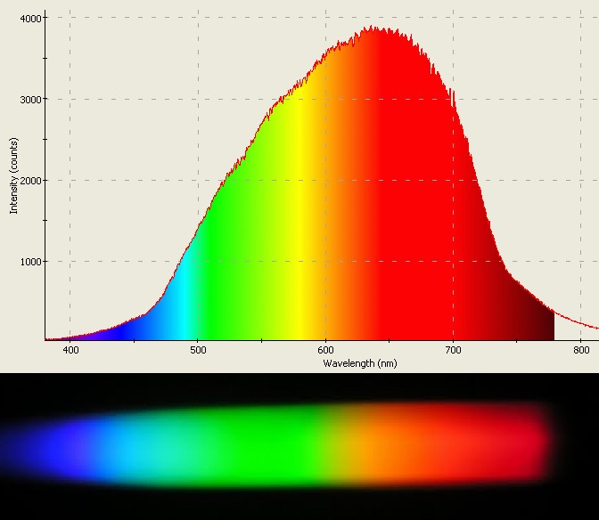
Smolina O. A., Engineer, smolinaoa@kolagmk.ru

Реферат

When studying the mineral composition of fine-grained powder samples and processing products, it may often be problematic to detect individual minerals due to the peculiar optical properties of fine particles in transmitted and reflected light. This primarily applies to rock-forming and low-reflecting ore minerals with the reflection of less than 10 %, for which no diagnostic features are available that could be established using polarizing reflection microscopes. The authors propose a new diagnostic technique based on the use of the optical parameter of «color temperature». This parameter is one of the fundamental characteristics of reflecting objects.Advanced photo and video cameras have the white balance function that controls the spectral composition of light in a microscope lamp. This enables measuring the «color temperature» of minerals in reflected light with conventional ore microscopes using the simplest video attachments for the eyepiece and a laptop. The «color temperature» is an important physical property of minerals that has not yet been used in mineralography. The new technique has been experimentally studied for a reference collection of approximately 20 minerals of apatite-nepheline and copper-nickel ores. The authors obtained stable parameters for determining the «color temperature» of minerals both in powders of artificial preparations and in ordinary polished thin sections for objects with the measured surface area of 30 μm2. The advantages of this technique include the rapidity and ease of mineral detection, which makes it a promising method for operational quality control of processing products.
The studies were carried out in the framework of Research Topic No. 0226-2019-0053 of the Geological Institute of the Kola Science Center of the Russian Academy of Sciences.

Ключевые слова Mineral detection, reflected light, color temperature, spectral composition of a microscope lamp, video attachment
Библиографический список

1. Methods of mineralogical research: Handbook. Ed. А. I. Ginzburg. Мoscow: Nedra, 1985. 480 p.
2. Agroskin A. S., Bezsmertnaya M. S., Vinogradova R. А., Zav’yalov Е. N., Lebedeva S. I., Papayan G. V., Petrov V. К., Rautian L. P., Sveshnikova О. L., Spiridonov E. М., Filimonova А. А., Chvileva Т. N. Guide-determinant of ore minerals in reflected light. Мoscow: Nedra, 1988. 504 p.
3. Bulakh А. G., Krivovichev V. G., Zolotarev А. А. General mineralogy. 4 ed., rev. and suppl. Мoscow: Akademiya, 2008. 416 p.
4. Neradovsky Yu. N. Ore mineragraphy. Murmansk: MSTU, 2009. 84 p.
5. Galopin R., Henry N. F. M. Microscopic study of opaque minerals. Cambridge: W. Heffer and Sons, 1972. 363 p.
6. Equipment catalogue. St. Petersburg: Analit, 2017. Iss. 3. 362 p. URL: http://analit-spb.ru/oborudovanie/potipu-oborudovaniya/ (accessed: 28.07.2019).
7. Focused ion beam — Scanning electron microscopy (FIB-SEM). Materials Science. Brno: TESCAM ORSAY HOLDING, a. s., 2015. pp. 18–20.
8. Nechiporenko А. P., Orekhova S. М., Plotnikova L. V., Glazacheva Е. N., Volkova К. V., Uspenskaya М. V. Specialized workshop on physical and chemical methods of analysis: electron and IR reflection spectroscopy, luminescent and x-ray fluorescence spectroscopy, refractometry, thermometry, kinetic pH-metry, indicator method — RCA. Theory and practice. Pt. II. St. Petersburg: ITMO, 2016. 181 p.
9. Melentev G. A., Shalygin V. A., Vorobjev L. E., Panevin V. Yu, Firsov D. A., Riuttanen L., Suihkonen S., Korotyeyev V. V., Lyaschuk Yu M., Kochelap V. A., Poroshin V. N. Interaction of surface plasmon polaritons in heavily doped GaN microstructures with terahertz radiation. Journal of Applied Physics. 2016. Vol. 119, Iss. 9. Paper 93104.
10. Wu J., Mayorov A. S., Wood C. D., Mistry D., Li L., Muchenje W., Rosamond M. C., Chen L., Linfield E. H., Davies A. G., Cunningham J. E. Excitation, detection, and electrostatic manipulation of terahertz-frequency range plasmons in a two-dimensional electron system. Scientific Reports. 2015. Vol. 5. Paper 15420.
11. Rasulov V. А., Ledeneva N. V., Iospa А. V Online diagnostics of clay-hydromicaceous mineralization based on diffuse reflection spectrometry in the range UV–VIS–NIR. Proc. of the Russian meeting with international participation «The role of technological mineralogy in rational subsoil use», Moscow, May 15–16, 2018. Мoscow: VIMS, 2018. pp. 162–165.
12. Egorova О. Е. Technical microscopy. 2 ed. Мoscow: Теkhnosfera, 2007. 360 p.
13. Optical microscope. URL: http://www.nikon-micro.ru/articles/292/ (accessed: 04.08.2019).
14. Chang F. H. A new X-ray diffraction method for quantitative multicomponent analysis. Advance in X-ray Analysis. 1973. Vol. 17. pp. 106–115.
15. Poole C. P., Farach H. A. Electron spin resonance of minerals. Part I. Nonsilicates. Magn. Reson. Review. 1977. Vol. 4. No. 3. pp. 137–195.
16. Poole C.P., Farach H.A. Electron spin resonance of minerals. Part II. Silicates. Magn. Reson. Review. 1978. Vol. 4. No. 4. pp. 225–289.
17. URL: https://a.d-cd.net/8c970cu-960.jpg (accessed: 28.07.2019).
18. Pozhilenko V. I., Gavrilenko B. V., Zhirov D. V., Zhabin S. V. Geology of ore districts in the Murmansk region. Apatity: КSC RAS, 2002. 359 p.

Language of full-text русский
Полный текст статьи Получить
Назад